
This startup’s technology could forever change our understanding of how the brain works, radically improving treatment options.
Anatomical brain mapping has been around since the last century, pioneered by innovators such as Korbinian Brodmann, who mapped out the whole human cerebral cortex over 100 years ago.
However, even modern-day brain maps are limited in scope, said Stephane Doyen, co-founder of Omniscient Neurotechnology.
“Most of the maps that exist are developed from the perfect brains of those available for research, such as young people,” he said. “But the brains doctors see have illnesses, such as tumours or previous surgeries that alter the shape of your brain.”
A former neuroscientist who left the field to pursue machine learning at a large-scale enterprise, Doyen knows this limitation all too well.
Having seen other industries use AI to improve operations, Doyen and co-founder Dr Michael Sughrue developed Omniscient in 2020 to bridge innovations in neuroscience with routine medical practice.
“Medical grade, neurosurgeon-supporting software needs to account for [different] brains,” he said. “So we had to find the data.”
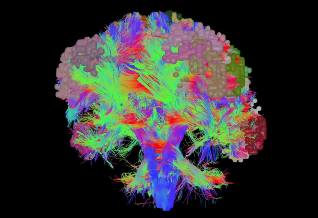
To do this, Omniscient’s team of engineers, data scientists and health-tech experts applied connectomics, which uses AI to look at the brain in terms of how it connects.
“We developed a map of the brain organising the cortex into a set of functional networks, which underpin specific functions of your brain,” said Doyen.
The start-up, one of a handful of consulting firm KPMG’s 2022 ‘Emerging Giants’, now has a staggering $400 million valuation, and TGA and FDA clearance to boot.
“We’re trying to make brain insights as available as possible so doctors and patients are empowered by knowing what’s going on in the brain,” said Doyen.
Mapping neural networks
Beyond just the imagery presented by a conventional medical scan, abnormalities can be identified by looking at the brain’s functional routes and connecting functional areas as a graph. This helps neuroscientists to measure things, said Doyen.
“When attempting to measure things previously, people were essentially putting together apples and pears, because they weren’t providing personalised answers,” he said.
For example, if you compare two different brains, the visual area – while in the same region – will be somewhat different.
“Therefore the measurement we’d be making for these specific points would be slightly off,” said Doyen.
Turning to MRI data for reference, which is more representative of the clinician’s patient subtype, Omniscient’s technology uses information gathered from a technique called Resting State BOLD (blood-oxygen-level dependent), which measures brain activity through oxygen levels in your blood.
“When your brain is firing, it sends a signal to the adjacent vessels, recruiting them to send more nutrients to the neurons,” said Doyen.
During an MRI, when patients are instructed to sit with no thoughts, their brains are still swirling with activity which can be represented by BOLD signals.
“When the brain goes into a resting state, the basic activity in the brain tends to show that things that work together, such as the neurons involved in vision, end up firing together – and this is how you can define networks,” he said.
“If you combine the map we have with that measure of activity, you now have a consistent way of describing the brain, meaning you can start to see clear patterns and draw comparisons between people.”
How it works
Omniscient’s star product is a cloud-based solution called Quicktome – targeted towards neurosurgeons today, but intended for all brain care specialists in the long term.
When a clinician decides that a patient needs an MRI, the data from that scan is transferred to hospital servers, which are then uploaded and processed through Quicktome’s cloud.
“We have a series of machine learning and data grooming processes in our cloud, as well as image-quality criteria assessments,” said Doyen.
Through this process, previously undiscovered patterns are inherited from brain scans at massive scales, enabling a personalised map of a patient’s brain to be accessible on a clinician’s own device.
“It’s a huge technological achievement to get there in under an hour,” said Doyen. “You can see which part of the brain is where, and an additional feature shows it’s functioning.”
Knowledge of patients’ functional neuroanatomy informs neurosurgeons how to approach a specific case.
For example, when a patient has a brain tumour, neurosurgeons might take the shortest pathway through the cortex to get to the tumour – but they may inadvertently cut through critical networks that are relevant for everyday life, such as speech or ability to move around.
“If you take a trajectory which is slightly more anterior or posterior and you don’t cut through those networks, you’ll have a potentially better outcome for the patient,” said Doyen.
Armed with better knowledge of where things are in the brain, this brings another level to informed consent.
“The patient knows a little bit better what’s ahead, and they can decide how far they are willing to go,” he said.
Other applications
Knowing where the functional areas of the brain sit also allows clinicians using Quicktome to identify other brain anomalies, including mental health conditions.
“Your behaviour, whether you’re feeling sad, happy, energised, tired or hungry, is all determined by networks in your brain,” said Doyen. “It’s also supported by a set of functional areas which are measurable.”
In the case of depression, your ability to anticipate rewards might be impaired.
“Using Quicktome, you can measure this function by looking at misfiring neurons and anomalies, projecting mental illness onto a biological level,” he said.
This enables better diagnostics by acknowledging that diseases have a constellation of symptoms.
“People who have depression might wake up early, have difficulty falling asleep, wake up in the middle of the night, or experience eating disorders,” said Doyen. “These are very different disease [presentations], so why should we use the same treatments?”
Empowering patients with the knowledge that “it’s not you, it’s your neurons” can provide significant relief.
“Patients can see, ‘I’m not sad all day because it’s my personality trait; it’s actually my brain which is misfiring’,” he said.
Another use for brain patterns in mental health is during patient follow up. “A lot of mental illnesses are chronic, [particularly] if you have them younger in life,” said Doyen.
“In follow up, you can see whether patients are developing the same patterns and take action before their symptoms progress and they have to undergo acute treatment.”
Looking forward, Quicktome could be used as a preventative technology to identify precursor signs for other brain conditions.
“We’ve published research about dementia, looking at specific impairments in hubs in the brain, which connect many functions,” said Doyen.
In the case of Alzheimer’s disease versus mild cognitive illnesses, Omniscient’s research identified that hubs are impacted in the former.
“That’s just one of the things we can see through connectomics, but there’s many more things we can look at as precursor signs.”