
Researchers have been trying to develop a biomaterial to patch the heart for more than a decade. Recent studies by engineers in the US could move things along.
In 2017, heart disease claimed 18,590 Australian lives, and almost 400,000 Australians have suffered a heart attack at some point in their lives.
Bioengineered cardiac patches have the potential to help improve heart function for heart attack survivors. But one challenge is making sure the engineered patch has a good network of tiny blood vessels – or microvessels – to make sure the patch survives and continues to work properly after it has been implanted.
To be effective, this microvessel network needs to be dense and highly aligned, like naturally occurring heart tissue. This depends on the design of the 3D scaffold used to grow the patch.
According to biomedical engineer Professor Feng Zhao, from Michigan Technological University in the US, the organisation of microvessels in 3D scaffolds has “largely been ignored” in previous research, which has focused on getting the cell alignment right.
“Understanding the mechanisms behind microvessel alignment in biomaterials will help us and other biomedical engineers to create better, more refined implants and devices,” Zhao explained in a statement.
Zhao and her team have developed a new biomaterial for cardiac patches that they hope will be a step in the right direction.
“The cardiac patch [developed] is completely biological, comprised of stem cells with vasculature that mimics real tissue, that could help repair a heart,” Zhao said.

Not too big, not too small – just right
According to Zhao, the heart presents some unique challenges for bioengineers.
“The cells are highly aligned for electromechanical signal transaction and the microvessels are also highly aligned and dense,” she explained.
Zhao recently published the results of an investigation into the pros and cons of six different bioengineering methods to create cardiac patches with highly aligned microvessels. These included 3D printing, surface patterning, microfluidics, micro-scaffolding, surface topography and electromechanical stimulation (stretching).
They found that none of the methods were able to reliably produce microvessel structures similar to human cardiac tissue. Bioengineered vessels had significantly larger diameters than the capillaries found in the heart, which would make it difficult for them to connect to the heart’s vessels and circulate blood when implanted. The engineered microvessel networks also tended to be less dense and mature than those found in natural tissue.
Some methods, such as 3D printing and microfabrication, rely on hydrogels, which isn’t able to match the strength or complex structure of natural heart tissue.
To find a solution to these issues, Zhao’s team took their cue from nature. In the body, cells are kept in position by a 3D structure made up of molecules such as collagen or enzymes. This is known as an extracellular matrix (ECM).
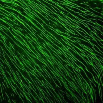
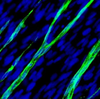
Using a 3D scaffold created by removing cells from a human ECM, the researchers were able to use adult human stem cells to construct a completely biological biomaterial with a blood vessel structure that mimicked real heart tissue.
The team’s research study detailing their method has been published as an advance online article in Theranostics. Now that they have successfully grown the biomaterial, the researchers will work towards clinical trials and refining their technique for use in implants and devices. And Zhao believes their method can be used to improve implants for other kinds of tissue.
“All tissues have this microstructure. It’s everywhere. This technology could be used for skeletal muscles, burn and chronic wound healing and nerve regeneration,” she said.