
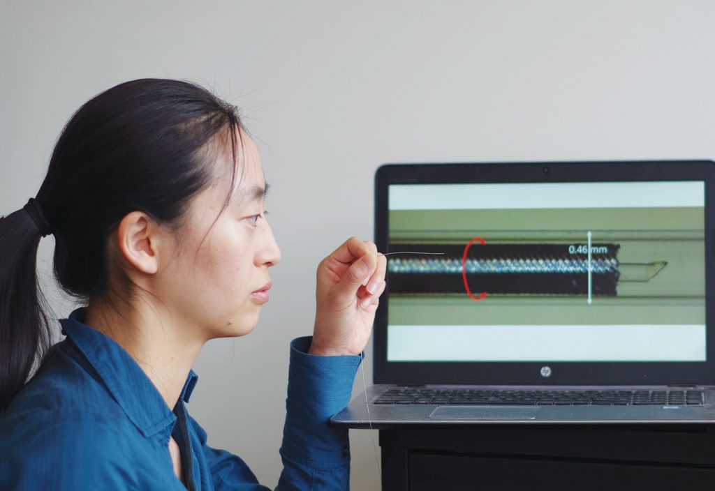
Biomedical engineer Dr Jiawen Li is developing ultra-thin fibre-optic devices that can explore the body’s organs and detect heart disease.
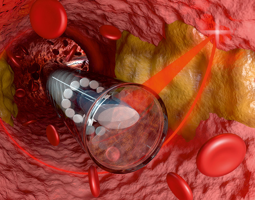
To provide clear images of plaque in the arteries that supply blood to the heart, an endoscope must be long and flexible enough to be inserted into the leg or arm, and able to be fed through the body’s arterial tree to the heart.
It must be thin enough to enter a partially blocked artery without causing trauma. And the picture quality must be high enough to visualise the plaque on the artery wall, measure the thin outer layer, and gather molecular information.
“There’s a lot of engineering involved,” said University of Adelaide biomedical engineer Dr Jiawen Li.
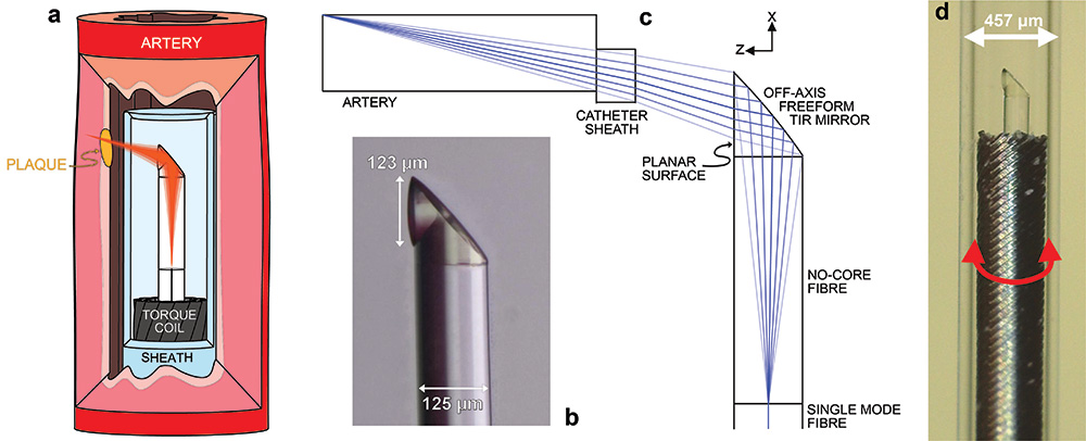
Li is developing next-generation medical devices that use optical coherence tomography, or “optical ultrasound”, to see inside the body’s organs and detect disease. She and her collaborators recently developed the world’s smallest freeform 3D-imaging catheter, which has a diameter of just 0.457 mm — including the protective outer tubing.

Hair’s breadth
Although optical ultrasound can be used to provide high-resolution, cross-sectional images of the body, it can’t see far into tissue.
“With regular biological tissue — like skin — we can only see to a depth of 1 mm to 2 mm,” said Li, who is funded by the National Heart Foundation.
“That’s why we need to use an optical fibre and build tiny endoscopes that can go deep into the body.”
Until now, image quality from these tiny endoscopic probes has been limited, reducing their usefulness in the clinic. But Li and her collaborators have developed a new fabrication technique that uses 3D microprinting to create an advanced, ultra-thin probe.
It contains side-facing micro-optics that are fewer than 130 μm in diameter — about the width of a human hair.
“What this device can also do is actually correct for all the nonchromatic optical aberration, which gives us better quality images,” Li said.
“All because it’s 3D printed.”
Li’s design was first made on a 3D printer so specialised there are only two in Australia. It uses two-photon polymerisation to produce freeform structures of nearly any 3D shape.
“When we print these miniature lenses on the end of an optical fibre, the features are tiny and the surfaces need to be incredibly smooth,” Li said. “Normal 3D printers don’t have the quality we need, but these two-photon printers have a spatial resolution of approximately 100 nm.
That’s smaller than the wavelength of the light we use to take the pictures.”
At the start of the research though, 3D printing caused more problems than it solved.
The printers lay down one layer at a time, and Li’s first design wasn’t smooth enough between the layers.
This caused the optical beam to diffract.
“At the very beginning, it was even worse than the traditionally made lens, which is polished,” Li said. “Polished lenses can actually be really smooth … but our first 3D-printed lenses were like a staircase.”

Li realised that some of the information in the design had been lost when it was being converted for the printer.
“When we do regular 3D printing it’s fine but when we go down to this nanometre scale, it becomes a big problem,” she said.
Li and her team overcame the challenge and successfully printed their first miniature lens for imaging blood vessels in 2018. But these early lenses still needed to be glued to the fibre.
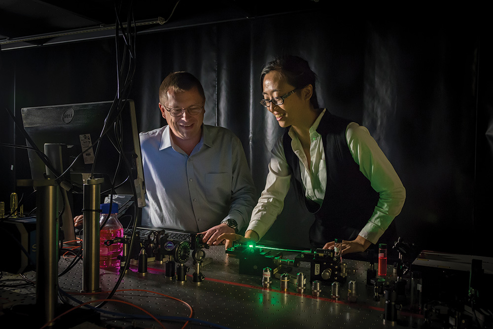
Later, Li collaborated with researchers at the University of Stuttgart, Germany, led by Professor Harald Giessen and Professor Alois Herkommer.
This team had developed a technique to 3D print directly on an optical fibre.
“Their innovation gave us the breakthrough we needed to make the world’s smallest endoscopic probe for heart disease,” Li said.
Medicine and technology
At the University of Adelaide, Li sits across both the Adelaide Medical School and the Institute for Photonics and Advanced Sensing.
“I’m often the one translating the medical questions into optical or engineering ones so that we can solve them,” she says.
One of the key features required for endoscopes is high resolution — something Li and her collaborators achieved by correcting for aberrations.
“It’s like putting a pair of glasses on to short-sighted eyes … everything becomes clearer,” she says.
The probe allows clinicians to visualise cholesterol crystals — tiny, crystallised structures that accumulate on the walls of blood vessels and are a significant risk factor for sudden cardiac arrest.
Head and heart
Coronary heart disease is the nation’s leading cause of death, killing more than 18,000 Australians in 2019.
If Li’s tiny endoscopes are successful, they could help doctors pinpoint high-risk plaque before it becomes life-threatening. But, she says, the technology could also be used to explore the nooks and crannies of other organs.
Li’s PhD research in the United States was driven by a desire to explore the brain after the death of her grandfather from a stroke.
“Inside the brain, there’s also blood vessels there, which, if they’re blocked, they will cause a stroke,” she said. “Those are areas that could be really useful for us to go in.”
Another potential application is pulmonary diseases and early-stage lung cancer. Being able to enter small airways could help doctors determine whether small tumours are malignant or not — something difficult to confirm from X-rays or CT scans.
Li has also been contacted by researchers interested in using the technology inside the ear.
“Our bodies are just filled with all those channels,” Li says.
“These endoscopes can go in different ducts, different vessels. And a lot of them would be really interesting to look into, not only for patients but also potentially for medical researchers.”