Engineers and scientists have combined their talents to flip traditional 3D printing and create intricate biomedical structures designed to regrow bones and tissues damaged by illness or injury.
The new method — which researchers have dubbed Negative Embodied Sacrificial Template 3D (NEST3D) printing — uses simple PVA glue as the basis for the 3D-printed mould.
Once the biocompatible material injected into the mould has set, the entire structure is placed in water to dissolve the glue, leaving just the cell-nurturing bioscaffold.
The aim of the emerging field of tissue engineering is to harness the body’s natural ability to heal itself, rebuild bone and muscle lost to tumours or injuries.
A key focus for biomedical engineers has been the design and development of 3D-printed scaffolds that can be implanted in the body to support cell regrowth. But making these structures small and complex enough for cells to thrive is a significant challenge.
Study first author PhD researcher Stephanie Doyle, who has a biomedical engineering degree from RMIT, said the advantage of the advanced injection moulding technique was its versatility.

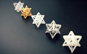
“We can produce dozens of trial bioscaffolds in a range of materials — from biodegradable polymers to hydrogels, silicones and ceramics — without the need for rigorous optimisation or specialist equipment,” she said.
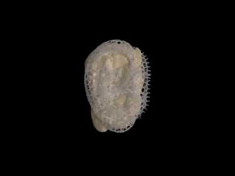
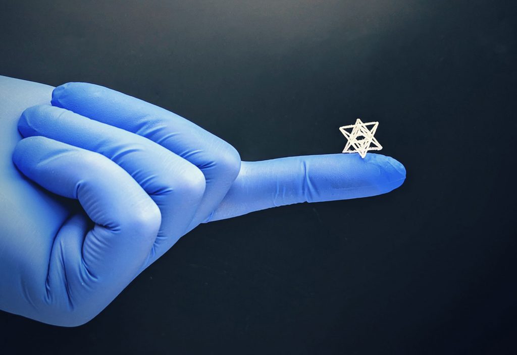
The 3D structures could be just 200 microns across, or the width of four human hairs, with complexity that rivals that achievable by light-based fabrication techniques.
“It could be a massive accelerator for bio-fabrication and tissue engineering research,” Doyle said.
The research team, led by Dr Cathal O’Connell, a Vice-Chancellor’s Postdoctoral Fellow at RMIT, collaborated with clinicians at St Vincent’s Hospital Melbourne, which houses Australia’s first collaborative, hospital-based biomedical engineering research centre.
These included orthopaedic surgeon Associate Professor Claudia Di Bella, cellular and molecular biologists Dr Serena Duchi, Dr Anita Quigley and Dr Carmine Onofrillo, and biomedical engineer Professor Elena Pirogova.
Future medical applications
O’Connell, a materials scientist, said it was extraordinary to create such complex shapes using a basic ‘high school’ grade 3D printer.
“That really lowers the bar for entry into the field, and brings us a significant step closer to making tissue engineering a medical reality,” he said.
The team had already created structures replicating human subchondral bone from a patient’s scan.
Doyle added that the NEST3D technique may also be applicable when looking at tissue engineering-based treatments for osteoporosis and arthritis.
“In fact, the immediate next stage of my work is focused on the repair of subchondral bone in an osteochondral knee defect in order to slow or prevent the onset of osteoarthritis,” she said.
“The NEST3D technique allows us to create a physical reinforcement structure out of a biological active material which, when combined with stem cells can provide the biological cues and environment to generate new bone tissue therefore filling the defect.”